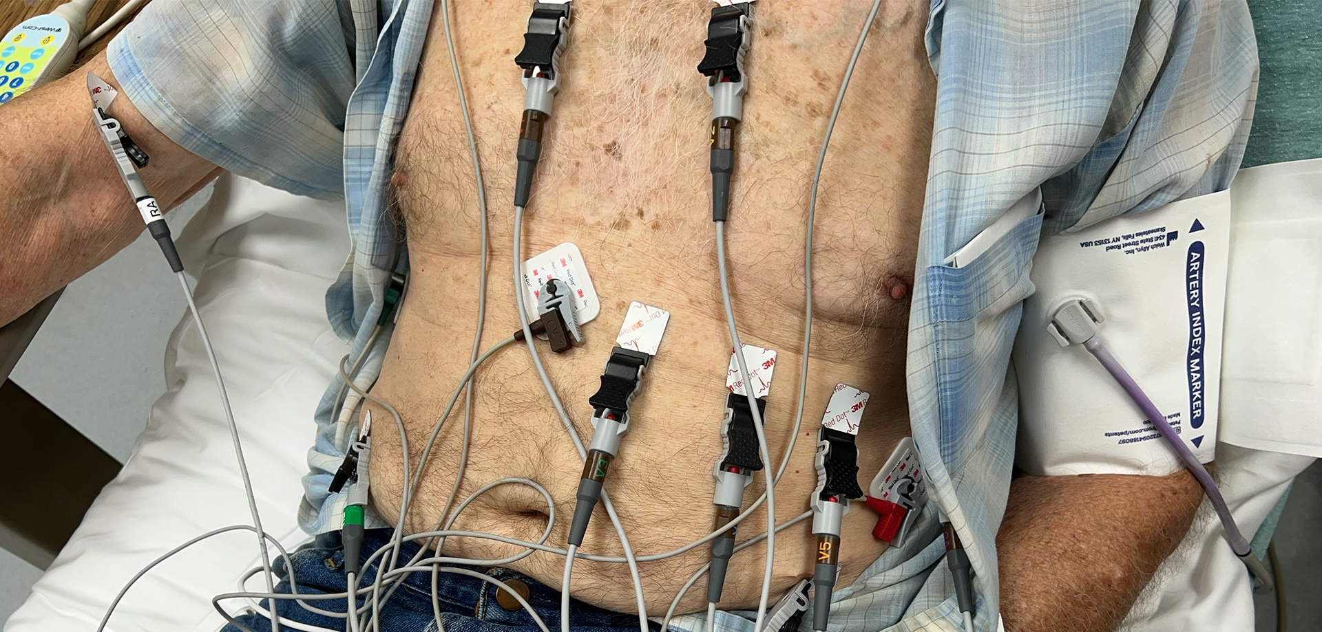
I saw a 51-year-old man recently with hypotension and tachycardia. It got interesting.
Continue readingAuthor: Jeffery Steele
Ultrasound Image Depth
Learners often make the same same mistakes. One of the most common is not setting the depth properly. It’s not hard to fix, but it does need to pointed out. Let’s do that.

Paced EKGs
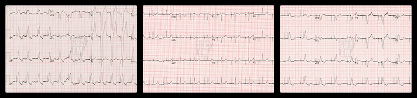
Not all “Paced” EKGs are the same. Let’s look at some of the differences among them.
Continue readingMonitor weirdness: concerning, yet oddly satisfying
There is nothing too deep here – and certainly nothing educational. These are just some vitals that caught my eye on the monitor.
Continue readingThe Cylinder Tangent Effect
Sometimes we measure cylindrical(ish) structures on a bedside ultrasound. On some occasions, the patient later gets a radiology performed scan and our measurements don’t match. This could be why.
Continue readingCervical spine badness… maybe?
I recently got some cervical spine X-rays on a patient that had been in a car wreck. You can probably guess what my level of concern was by virtue of the fact that I did not get a CT. I did, however, find something interesting…

X-rays or Ultrasounds for fractures?
Bones are very echogenic, which makes their ultrasound appearance a bright white line that is often easy to see – particularly in the case of superficial long bones. That’s what makes ultrasound a great way to find fractures. Let’s look at some examples.
Continue readingWhat’s the heart rate: follow up
In the last post, I mentioned that I was not sure why there was no palpable pulse from the PVC during ventricular bigeminy. Does the PVC not cause a mechanical contraction at all? Or does it actually cause a contraction that is too weak to feel? I’d mentioned that I could have answered the question with an ultrasound.
It was just a matter of time before this happened again.
Continue readingWhat’s the heart rate?
Here we see a nice example of ventricular bigeminy. Besides the rhythm, do you notice anything strange on the cardiac monitor?