Gall stones within the body or fundus of the gall bladder are usually pretty easy to find on point of care ultrasound. There is, however, a scenario in which they can be hidden. We have to be diligent and avoid some pitfalls to find those.
It takes most learners five to ten scans to reliably be able to find the gall bladder, and the quality of the images improves with repetition and guidance thereafter.
In one frequently referenced study, after 25 scans most learners could “identify gall stones.” My experience working with learners certainly agrees with that number for being able to reliably identify stones in the body or fundus of the gall bladder. Several characteristics make those stones some of the lowest-hanging fruit in all of ultrasound. Specifically, their dense composition reflects all of the ultrasound beam back to the transducer, which results in a bright white (hyper-echoic) appearance with a dense shadow deep to it. Also, the surrounding bile transmits all of the ultrasound beam creating a wonderful “acoustic window” of black space to make their white surfaces distinct and hard to miss.
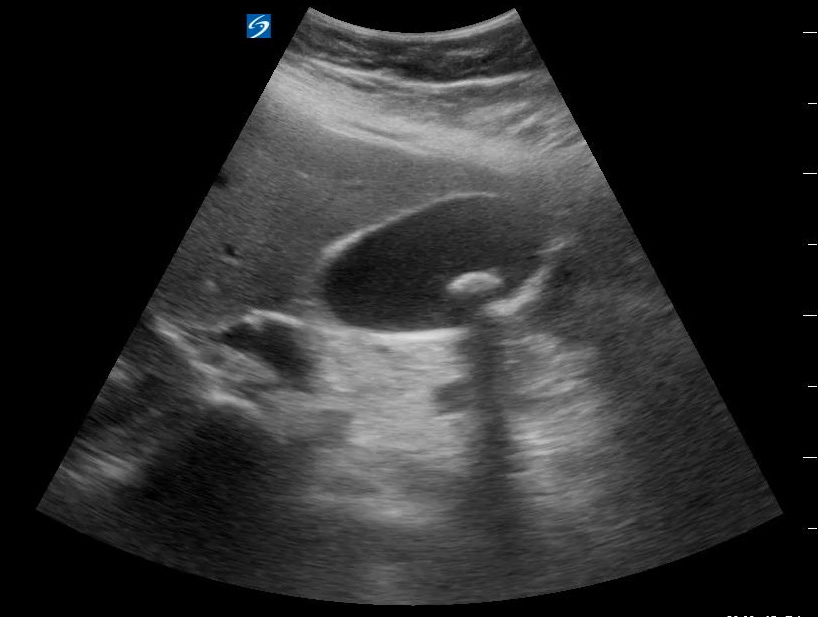
Unfortunately, when the stone is in the cystic duct or deep within the neck of the gall bladder, they do not stand out as prominently. The lack of surrounding bile and the presence of other sources of shadows make stones in these deeper locations a challenge to identify.
Example 1
Here we see a long axis view of a gall bladder with no stones visible in the body nor fundus. It remains suspicious, however, as it is distended and has a diffusely thickened wall.
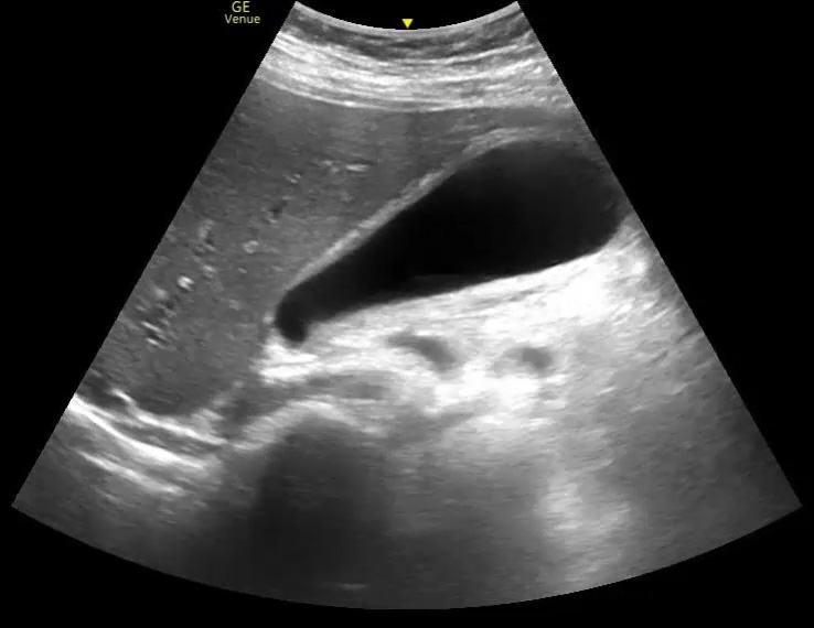
This is the same gall bladder as above. Scrutinizing the neck, however, has revealed a stone that blends in with the surrounding structures. The shadow in this case is what really catches the eye. This is a stone in the cystic duct. Notice that although the stone does have that characteristic hyper-echoic (bright white) appearance, outside the body of the gall bladder there are many other similarly colored structures. Stones in the cystic duct can be camouflaged by the surrounding tissue, and the shadow may be the best (or only) clue to its presence.
Example 2
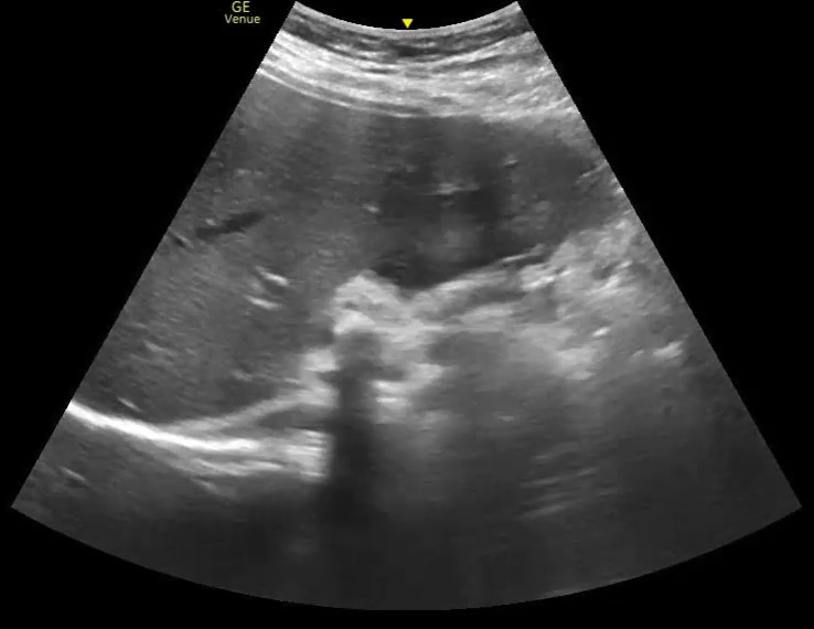
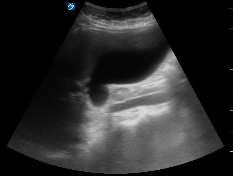
Here we see another distended gall bladder without stones in its body.
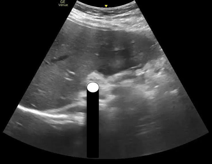
Moving the porta hepatis to the middle of the screen and scrutinizing it reveals a stone in the cystic duct that had not been visible on the prior scan. Again, the shadow is prominent, though the stone itself could easily have been overlooked as there are other hyper-echoic structures in the area.
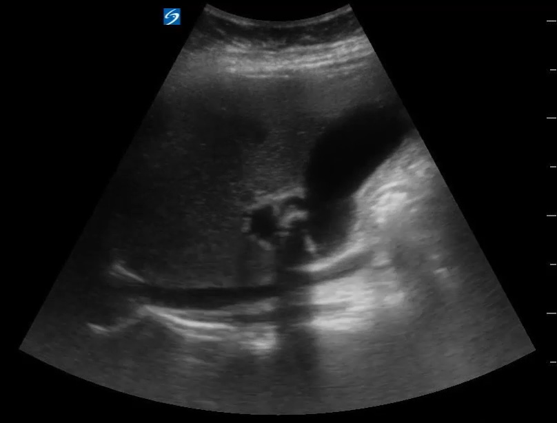
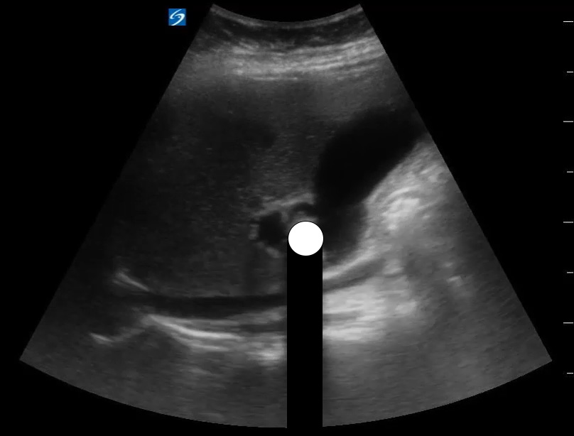
These two scans are perfect examples of times when there are no stones in the gall bladder body, but there are stones in the cystic duct. Those stones could easily have been overlooked if we hadn’t taken the time to really focus on and scrutinize the neck of those gall bladders. They can be very humbling.
As we have seen, without the acoustic window provided by the bile in the gall bladder, sometimes the acoustic shadow of the stone is our best bet for finding one in the cystic duct. Unfortunately, there are often lots of shadows in the area around the gall bladder. Some are due to bowel gas, but others are due to an imaging artifact called edge artifact. These can get in the way and lead to misdiagnosis.
Edge Artifact
Edge artifacts are dark, shadow-like lines that occur at the edges of curved, smooth walled structures like gall bladders, blood vessels, or the urinary bladder. They are due to bending of the ultrasound waves as they bounce off of those structures. That bending causes less than the “expected” amount of waves to return to the transducer. The computer is therefore “tricked” into thinking there is nothing at that depth, which it displays as a black line on the screen.
That is a little different than true acoustic shadowing. Acoustic shadowing occurs when a structure that is opaque to the ultrasound beam blocks all transmission. The area on the screen deep to the opaque object is “invisible” to to the transducer and therefore displayed as black on the screen. Acoustic shadowing is not an issue of the transducer getting “fooled” into misrepresenting what is actually there as is the case with edge artifact.
When looking for gall stones, noticing the shadow they cast is a common, indirect way to find them. Having other “shadows” in the image could make us think there is a gall stone present that is not – a false positive. Or, they could obscure a stone that does exist – a false negative.
Recognizing edge artifact and how is differs from acoustic shadowing can help you avoid those pitfalls when looking for stones that are not in the body of the gall bladder.
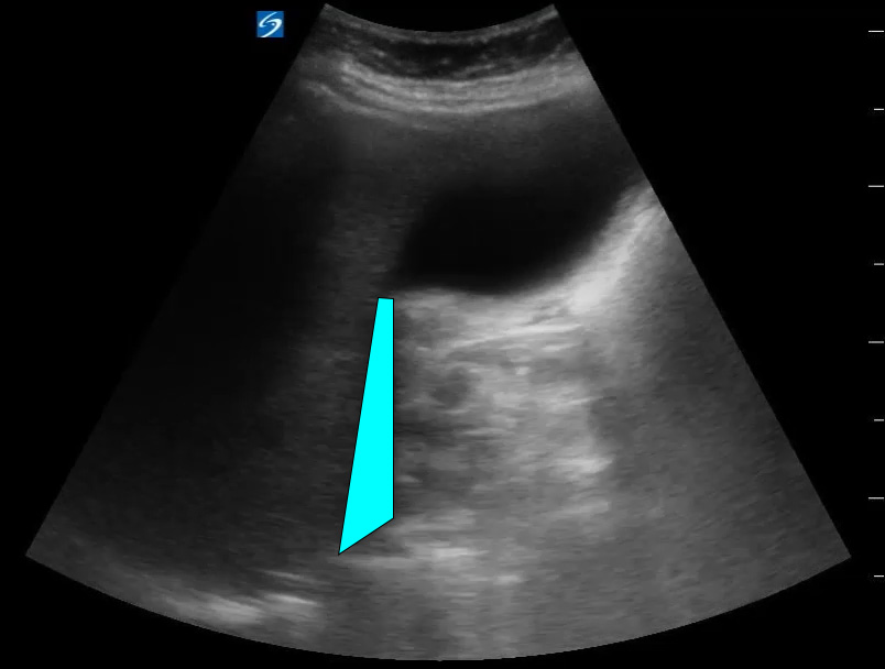
Example 1
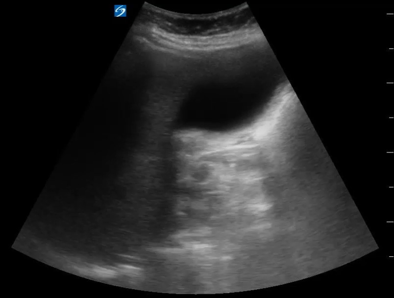
Notice the shadow on the left side of the screen at the edge of the gall bladder.
Example 2
This gall bladder has a bend that generate edge artifact from more than one of its surfaces.
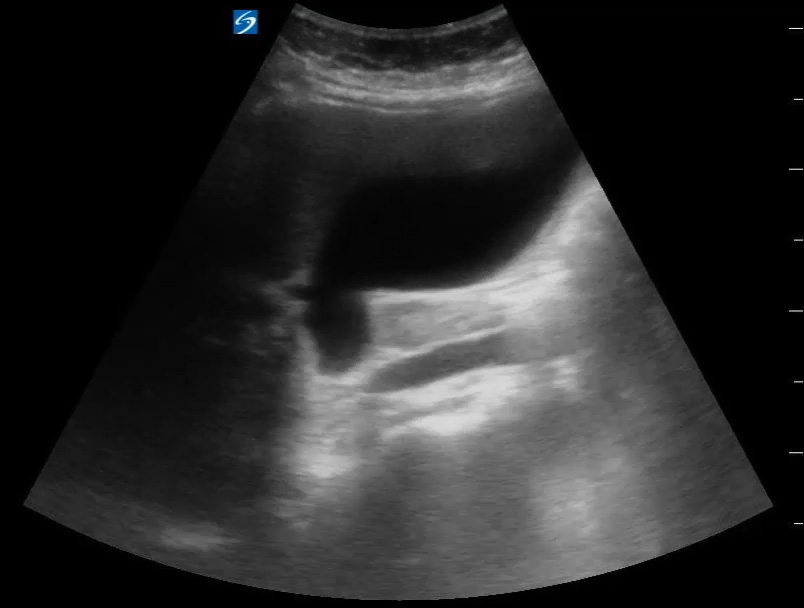
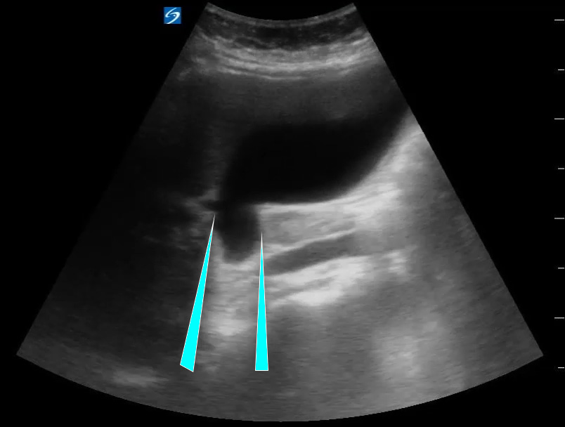
Cases like these serve as a reminder of the fact that most of us doing point of care ultrasound are doing “limited” studies. In the case of gall bladders, the thing we expect to reliably be able to find is gall stones in the body. More subtle findings like stones outside of the gall bladder as well as secondary signs of cholecystitis (wall thickening, free fluid, ductal dilation) are a step beyond simply the presence or absence of gall stones. Those skills can be obtained with practice and humility, but be aware of where your limitations are and what you can truly rule in or out with the skill set that you possess. Who knows how many of these I’ve missed over the years.
Scan happy, my friends.

Reference
Gaspari RJ, Dickman E, Blehar D. Learning curve of bedside ultrasound of the gallbladder. J Emerg Med. 2009 Jul;37(1):51-6. doi: 10.1016/j.jemermed.2007.10.070. Epub 2008 Apr 25. PMID: 18439787.

