Cardiology
Ventricular Tachycardia and adenosine
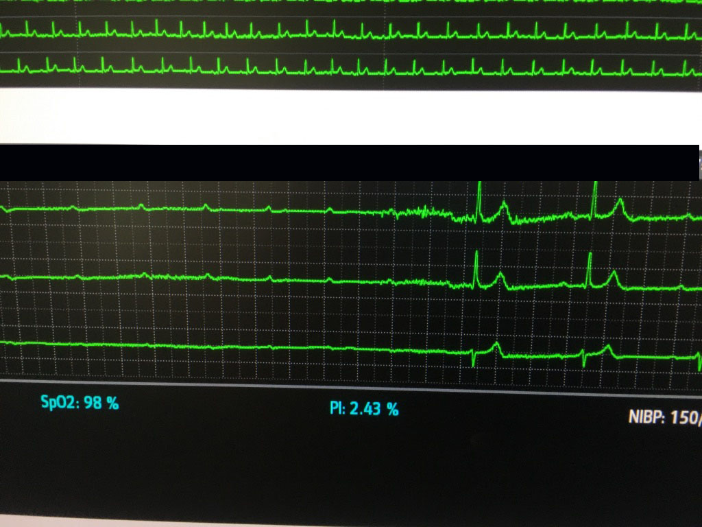
Supraventricular Tachycardia with abnormal conduction (“aberrancy”) is often difficult to distinguish from Ventricular Tachycardia. The 2015 AHA guidelines on the management of adults with SVT state that the presence of AV dissociation (i.e., the presence of P waves visible among the QRS complexes at a rate slower than the ventricular rate) or fusion/capture beats “provides the diagnosis of ventricular tachycardia.” Other criteria are suggestive, but not confirmatory. Diagnostic algorithms – Brugada or Vereckei for example, are complicated and can be difficult to apply.
Continue readingSTEMI
The formal definitions of a STEMI are the following:
New J point elevation, in the absence of LBBB and LVH of:
- Greater than or equal to 1 mm in at least 2 contiguous leads EXCEPT leads V2 and V3
- Leads V2-V3 require:
- greater than or equal to 2.5 mm in men less than 40
- greater than or equal to 2 mm in men over 40
- greater than or equal to 1.5 mm in women
- Inferobasal STEMI’s (previously known as posterior infarcts):
- Isolated ST depression greater than or equal to 0.5 mm in leads V1-V3
- especially when the terminal T wave is positive
- If these are present, STEMI is confirmed by STE in posteror leads (V7-V9), which should be obtained:
- STE greater than or equal to 0.5 mm or
- STE greater than or equal to 1 mm in men < 40
- Reference:
- Govea A, et al. Interv Cardiol Clin. 2021 Jul;10(3):293:306.
