Indulge me in a few more thoughts and images about posterior vitreous detachments.
Where did we leave off?
In the prior post about retinal detachments we established a few things:
- Retinal detachments can look similar to vitreous detachments on ultrasound.
- Of the two, retinal detachments are more concerning as some may benefit from an urgent specialist evaluation.
- Retinal detachments can be distinguished from vitreous detachments by the fact that the retina is an extension of the optic nerve. Retinal detachments will therefore always be tethered to the optic nerve. To that end, it is imperative that your scan include the optic nerve.
- I did not have an example of a vitreous detachment, because they are generally not symptomatic other than flashes and floaters and aren’t the type of thing people come to an emergency department for.
It bothered me that I didn’t have an example
I made a point to look for a vitreous detachment. Supposedly it s a common thing that occurs as we age, so it should not be that hard to come across one, right?
It took less two weeks to find a few great examples, and they bring up a VERY IMPORTANT point that I’d not put much thought into before: the gain setting.
The following clips are from the same patient and were done at the same time.
Clip 1: default ocular ultrasound gain setting
This is a benign looking exam. Nothing to comment on. This is what eyes are supposed to look like.
Clip 2: same eye as clip one, but with the gain turned all the way up
Notice what a difference the gain setting makes. There is clearly a vitreous detachment that was completely invisible at the default gain setting
Retinal detachments, to the contrary, will be visible at standard gain settings:
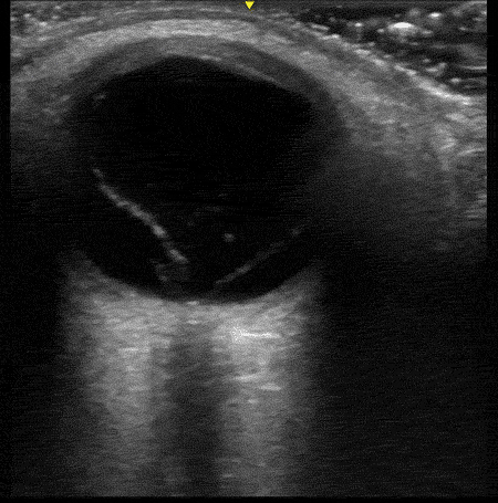
[You can tell the gain is set very differently here than the clip with vitreous detachment because of how bright the image is. It is particularly noticeable in the part of the image deep to the globe due to an ultrasound artifact called “posterior acoustic enhancement”; but I digress.]
The learning point is that you will almost certainly have to turn the gain up see vitreous pathology. In fact, increasing the gain often reveals things floating around in the vitreous humor that you would never have seen or known about – “mobile vitreous opacities.” These things have little (if any) clinical significance, and you’re probably just as well off not to have seen them.
More vitreous fun
Clip 3: same person as clips one and two, same time, other eye; default gain setting
Clip 4: Same eye as clip three, gain turned all the way up
Once again, clear vitreous detachment that was completely invisible at standard gain settings.
This person was in their late sixties and has some degree of vitreous detachment in both eyes. They said they very occasionally notices floaters, but otherwise do not have symptoms from the pathology.
The learning point of the second set of clips is appreciating how a vitreous detachment moves when the person looks from side to side. It “sloshes”. It keep rolling after the eye stops moving akin to clothes in a dryer. Compare that to the retinal detachment example above. The characteristics of the movement can be used to help distinguish the two.
Wrap it up
Posterior vitreous detachments are out there and can have a similar appearances to retinal detachments. Unlike retinal detachments, however, vitreous detachments are not tethered to the optic nerve and will be very difficult to see at standard gain settings. They also move differently when the eye rotated.
I find it reassuring that that there are multiple ways to keep from making the mistake of confusing a potentially emergent diagnosis with a relatively benign one.
Scan happy, my friends
