I recently got some cervical spine X-rays on a patient that had been in a car wreck. You can probably guess what my level of concern was by virtue of the fact that I did not get a CT. I did, however, find something interesting…

The films
Wait a darn minute…
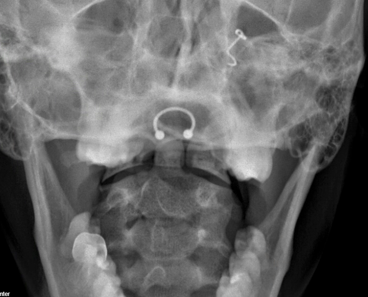
A close look at the odontoid view showed something concerning.


There is a line at the base of the dens concerning for a type II fracture, which would be considered unstable.

(Here is a link to radiopaedia’s page on odontoid process fractures if you need a refresher.)
Something didn’t fit, though. This patient’s wreck had been a week ago. Also, the line feels a little “too clean”. I looked at it with the residents, but the answer was not clear to them. Do you see any issues?
A hint…
I’ll highlight two structures. Do you see it now?
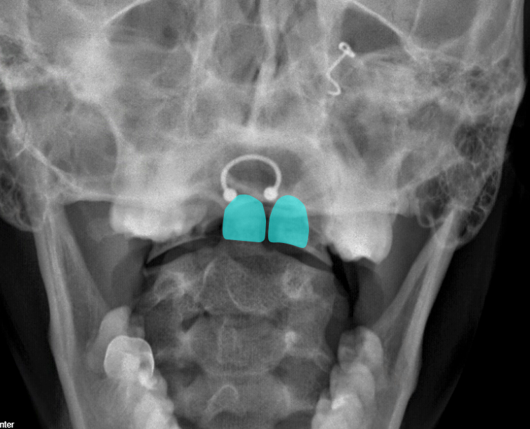
The line was an overlap of one of the incisors – a so-called “tooth artifact”.
A word about the adequacy of the films
Other than motion artifact, we hardly ever have “inadequate” CT scans. That’s not the case with X-rays where positioning is so important.
For the open mouth view in a cervical spine series to be “adequate,” we need to see the entire dens and the lateral masses of C1 as they articulate with C2. In this case, we can’t really see the entire dens and what we can see is obscured by teeth. We can see the most inferior bit of the lateral masses, enough to see normal alignment, but we can’t really see any of the body of C1.
Either this patient didn’t open their mouth wide enough, or the image wasn’t shot at a steep enough angle to get the teeth out of the way.
Since we are mentioning adequacy of images, lets invest a moment on the lateral.


To be considered adequate for interpretation, we need to see the articulation of C7 on T1, which unfortunately we don’t here. So, again, this study leaves something to be desired.
This case was fun example of time when a common artifact could have sent us down the wrong diagnostic path. As they say, “the eye does not see what the mind does not know.” Now your mind knows this one.
A Lucky Coincidence
This was actually rather timely. In July of 2024, the Pediatric Emergency Care Applied Research Network (PECARN) published an observational study and prediction rule for cervical spine imaging in children with blunt trauma. In it they recommend plain films for a subset of patients. Although this prediction rule has not been adopted into practice at this point, the idea that some children do need imaging and that plain films are adequate in at least some of them could be a very positive thing.
That being said, if the rule pushes you to get plain films of patients in whom you would have otherwise done no imaging, that’s a step backwards. On the other hand, if your practice has been “CT or bust” and this sheds light on who can have the cheaper, quicker, less radiation images without compromising safety, that would be a win. If that’s the case, we may be ordering a few more plain films of cervical spines in the coming years. If so, we will see more of this sort of artifact that make the films occasionally difficult to interpret – a fact that probably contributes to why it is often so tempting to go straight to CT in current times.
TOOTH ARTIFACT. How about that?
References
Leonard JC, Harding M, Cook LJ, Leonard JR, Adelgais KM, Ahmad FA, Browne LR, Burger RK, Chaudhari PP, Corwin DJ, Glomb NW, Lee LK, Owusu-Ansah S, Riney LC, Rogers AJ, Rubalcava DM, Sapien RE, Szadkowski MA, Tzimenatos L, Ward CE, Yen K, Kuppermann N. PECARN prediction rule for cervical spine imaging of children presenting to the emergency department with blunt trauma: a multicentre prospective observational study. Lancet Child Adolesc Health. 2024 Jul;8(7):482-490. doi: 10.1016/S2352-4642(24)00104-4. Epub 2024 Jun 4. PMID: 38843852; PMCID: PMC11261431.
