Bones are very echogenic, which makes their ultrasound appearance a bright white line that is often easy to see – particularly in the case of superficial long bones. That’s what makes ultrasound a great way to find fractures. Let’s look at some examples.
Case 1
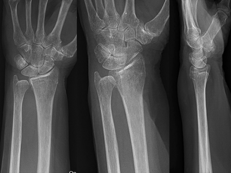
Here we see long axis views of the distal radius and ulna in a 12-year-old with a FOOSH injury. The fracture is as plain as day – even the ulnar styloid fracture. In this case, we took the ultrasound machine into the room during the initial evaluation and had these images before we left the bedside.


Case 2
Here we see a long axis view of a distal radius on a 79-year-old lady with a fall. In this case, there is a non-displaced fracture, which is actually more clear on the ultrasound than the X-ray. This fracture is clearly not angulated.
Case 3
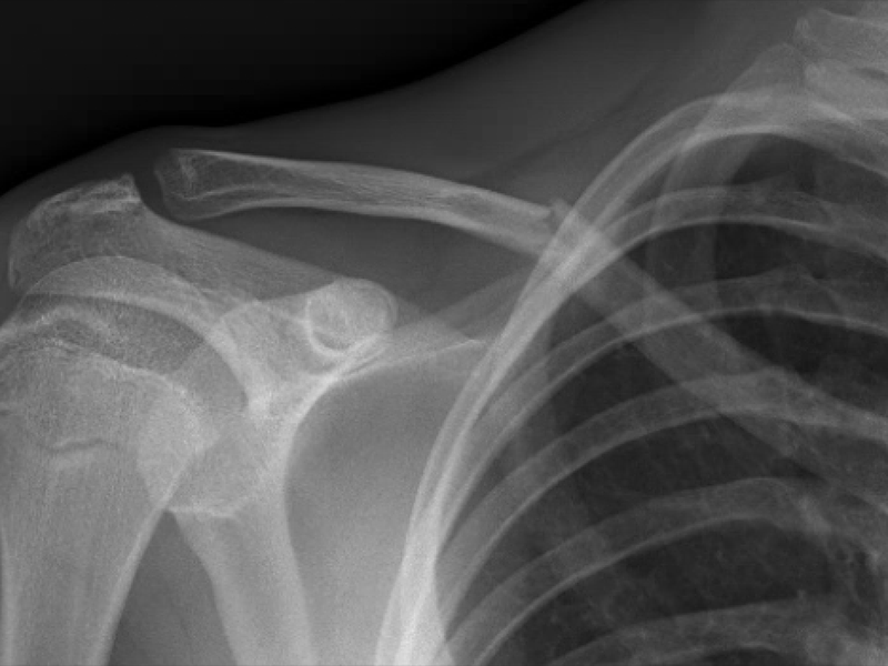
Here we see a 17-year-old football player with a shoulder injury. Again, we had the answer before we left the bedside as the displaced fracture is clear on the ultrasound.


There are studies – we did one at journal club recently – showing that point-of-care ultrasound can be used in this fashion, and these are nice examples of how clear fractures can be on superficial long bones.
Alas, we still got the X-rays. The orthopedic surgeon is going want them. There’s no way around it – at least not at my shop at the present time.
As much as I like doing them, I have to wonder if this a solution to problem that’s not really a problem.
When would this be helpful?
I can think of a few times.
- I do use this when I reduce fractures. I like a “before” scan, then a quick look once I think I have anatomic alignment for confirmation before I go to the trouble of putting the splint on. I have found this incredibly helpful. It’s sort of a poor man’s C-arm.
- If time permits, I like having a quick look while we are waiting on the X-ray. I can go ahead and make my plan. Also, the patients and their families seem to like seeing the images, and the face time with the provider is always a crowd pleaser.
Bottom line
I look at these as fun tests that patients enjoy, which are good for ruling in fractures. However, I admit that I don’t routinely do a thorough enough exam to rule out all fractures or bony pathology. I could be more meticulous if X-rays weren’t available, but they always are. At this moment in my life, I find it’s most practical use is in fracture reduction as a make-shift C-arm.
Scan happy, my friends

Reference
- Snelling PJ, Jones P, Bade D, Bindra R, Byrnes J, Davison M, George S, Moore M, Keijzers G, Ware RS; BUCKLED Trial Group. Ultrasonography or Radiography for Suspected Pediatric Distal Forearm Fractures. N Engl J Med. 2023 Jun 1;388(22):2049-2057. doi: 10.1056/NEJMoa2213883. PMID: 37256975.
