A thrombus in the heart is really the holy grail of point-of-care ultrasound. Here are two great examples.
Left Ventricle Thrombus
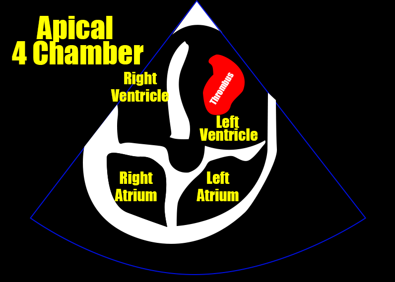
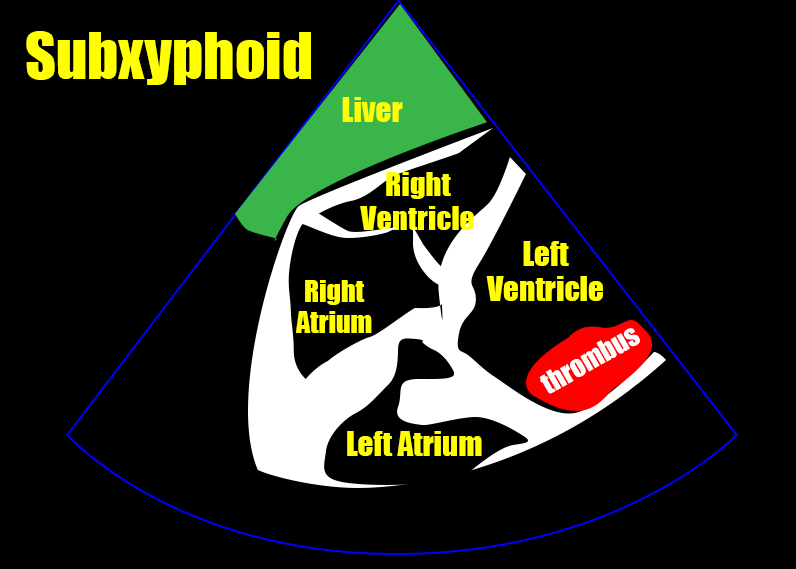
The following three images show a clot in the left ventricle. Notice that it adheres to the ventricular wall. Such clots are called mural thrombi. They form within the ventricle itself as opposed to having formed elsewhere and embolized. They are the result of disruptions to laminar blood flow through the heart. Common causes include dilated cardiomyopthies, post ischemic regional wall motion abnormalities, atrial fibrillation, or very low ejection fractions. As the left side of the heart is emptied by the aorta, left ventricular thrombi pose a risk for peripheral arterial occlusions – specifically embolic strokes. However, if the clot embolized through the aorta but did not make it up the carotids, other peripheral infractions could certainly arise as well.
Of note, a right sided clot could get to the left ventricle if there were a ventricular septal defect or other communication between the two sides. However, that would not cause a mural thrombus, as we see here.


Right Atrial Thrombus
The following two images show clot moving back and forth between the right atrium and the right ventricle. These are free-floating and are often referred to as thrombus-in-transit or clot-in-transit. Unlike mural thrombi these do not form in the heart itself but rather have embolized from a venous thrombus. As the right side of the heart empties by way of the pulmonary artery, these are destined to become pulmonary emboli. The mortality rate of this finding is very high, usually said to be greater than 25%. If you find this, decisions will have to be made about an attempt of embolectomy, anti-coagulation, or thrombolytic therapy.
Notice how large the right ventricle is on the apical four chamber view. A normal right ventricle is ~ 2/3 the size of the left ventricle. In this case, however, the right is considerably larger than the left. In this setting, this finding tells us that the clot in the right atrium is not the first one. This person already has a pulmonary embolism large enough to cause right heart strain.


That’s it. These are interesting and uncommon images the likes of which I’d never seen before a few months ago. I truly feel like we have found the grail.

