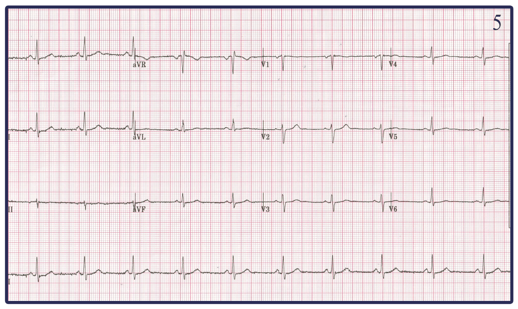
Here we see 5 electrocardiograms. All were done on the same person (a brilliant and handsome 41 year old EM physician) at the same time. There are clearly differences among them. One of them is ‘correct.’ To get you started, these are lead reversals.
- Which one is correct?
- Which of the others represents which lead reversals.
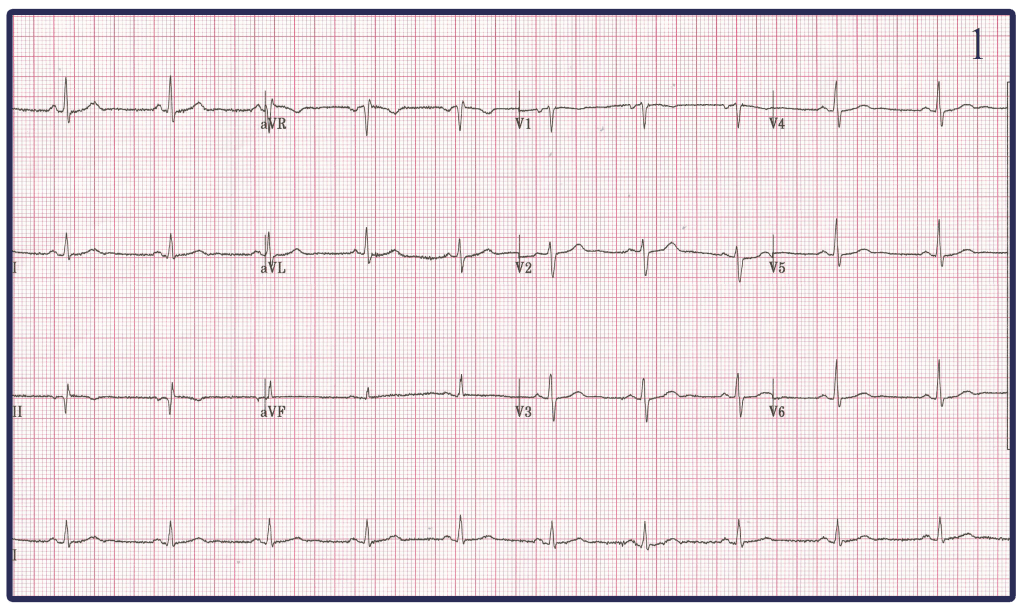
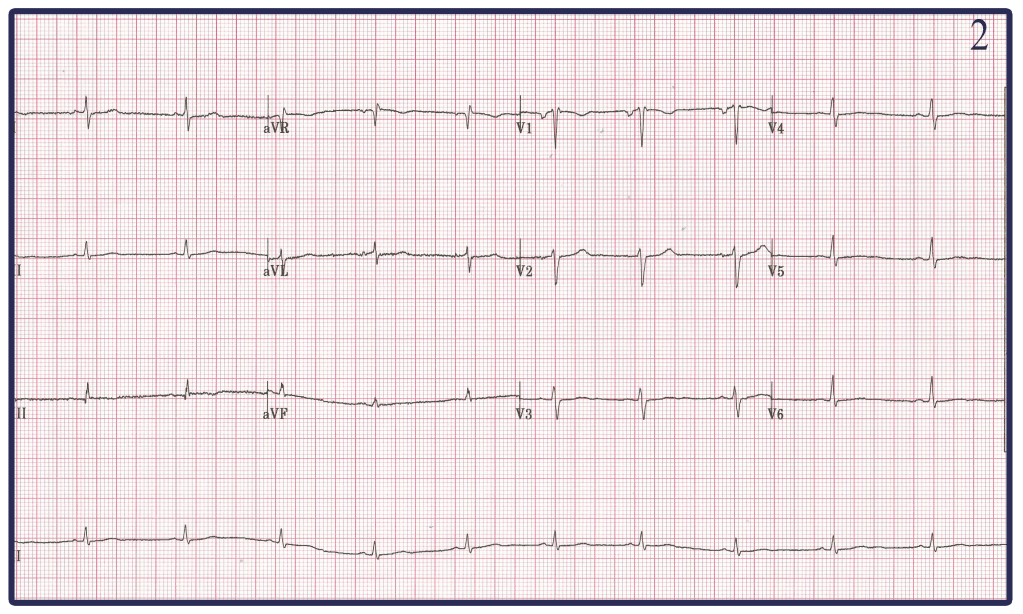
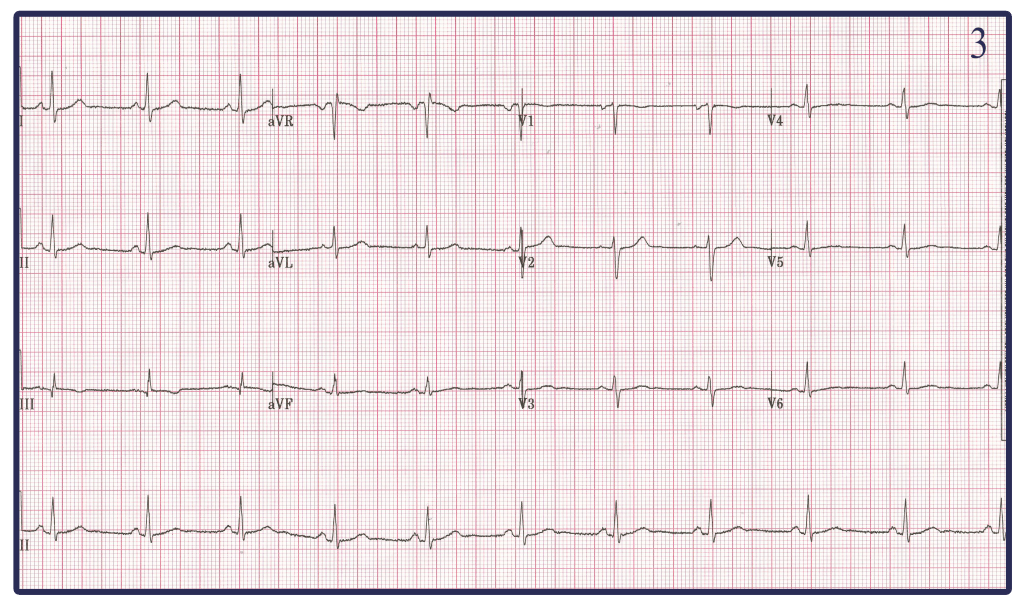
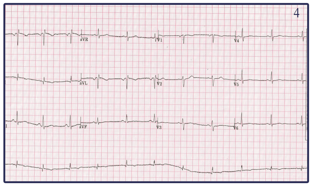

Based on the Einthoven Triangle my answers are below. Whether or not they are correct I have no idea.
-I think EKG 3 is the correct one based on Leads I, II, and III being postive and aVR having negative p waves and normal R wave progressions in the chest leads.
-EKG 4 looks like a reversal in the RA and LA leads causing leads II and III to switch places as well as leads aVR and aVL to switch places when compared to the normal EKG 3.
-EKG 5 is reversal in leads LA and LL because when compared to EKG 3 leads aVF and aVL switch places and leads I and II switched places but aVR is unchanged.
-Both EKG 1 and 2 are more of a mystery to me. Honestly looking at EKG 2 and it appearing to be low voltage I would have assumed this to be caused by someone Obese. However I happen to know this 41 year old EM Physician (beauty is in the eye of the beholder) and would not consider him Obese. My best guess is that one of the leads in EKG 1 and EKG 2 was exchanged with the negative terminal RL changing the morphology of not only the Limb leads but also the chest leads in both.
LikeLike
3 is correct.
1- leads III & V6
2- leads II & aVF
4- leads aVR & aVL
5- leads aVL & aVF
LikeLike
So, I’d like to retract my previous statement. My brain truly was busted.
1. RL & RA
2. RA & LL
3. Correct
4. LA & RA
5. LA & LL
LikeLike